
A biological membrane, biomembrane or cell membrane is a selectively permeable membrane that separates the interior of a cell from the external environment or creates intracellular compartments by serving as a boundary between one part of the cell and another. Biological membranes, in the form of eukaryotic cell membranes, consist of a phospholipid bilayer with embedded, integral and peripheral proteins used in communication and transportation of chemicals and ions. The bulk of lipids in a cell membrane provides a fluid matrix for proteins to rotate and laterally diffuse for physiological functioning. Proteins are adapted to high membrane fluidity environment of the lipid bilayer with the presence of an annular lipid shell, consisting of lipid molecules bound tightly to the surface of integral membrane proteins. The cell membranes are different from the isolating tissues formed by layers of cells, such as mucous membranes, basement membranes, and serous membranes.
YouTube Encyclopedic
-
1/5Views:4 840 5033 229 4891 753 828131 178481 098
-
In Da Club - Membranes & Transport: Crash Course Biology #5
-
Cell Transport
-
Inside the Cell Membrane
-
Cell Biology | Passive & Active Transport | Endocytosis & Exocytosis
-
Structure Of The Cell Membrane - Active and Passive Transport
Transcription
Oh, hey! I didn't see you up there. How long have you been waiting in this line? I've been here for like 15 minutes and it's freaking freezing out here I mean, whose banana do you gotta peel in order to get into this club? Well, while we're here I guess this might not be a bad time to continue our discussion about cells. Because cells, like nightclubs, have to be selectively permeable. They can only work if they let in the stuff that they need and they kick out the stuff that they don't need like trash and ridiculously drunk people and Justin Bieber fans. No matter what stuff it is it has to pass through the cell's membrane. Some things can pass really easily into cells without a lot of help, like water or oxygen. But a lot of other things that they need, like sugar, other nutrients, signaling molecules or steroids they can't get in or it will take a really long time for them to do it. Yeah. I can relate. Today we're going to be talking about how substances move through cell membranes, which is happening all the time, including right now, in me and right now, in you. And this is vital to all life, because it's not just how cells acquire what they need and get rid of what they don't, it's also how cells communicate with one another. Different materials have different ways of crossing the cell membrane. And there are basically two categories of ways: there's active transport and there's passive transport. Passive transport doesn't require any energy, which is great, because important things like oxygen and water can use this to get into cells really easily. And they do this through what we call diffusion. Let's say I'm finally in this show, and I'm in the show with my brother John. Some of you know my brother John, and I love him, but he uh... He's not a big fan of people. I mean he likes people. He doesn't like big crowds. Being parts of big crowds and people standing nearby him, breathing on him, touching him accidentally and that sort of thing Because John's with me at the show, we're hanging out with all of our friends near the stage. But then he starts moving further and further from the stage so he doesn't get a bunch of hipsters invading his space. That's basically what diffusion is. If everyone in the club were John Green they would try and get as much space between all of them as possible until it was a uniform mass of John Greens throughout the club. When oxygen gets crowded, it finds places that are less crowded and moves into those spaces. When water gets crowded, it does the same thing and moves to where there is less water. When water does this across a membrane, it's a kind of diffusion called osmosis. This is how your cells regulate their water content. Not only does this apply to water itself, which as we've discussed is the world's best solvent. You're going to learn more about water in our water episode. It also works with water that contains dissolved materials, or solutions, like salt water, or sugar water, or booze, which is just a solution of ethanol in water. If the concentration of a solution is higher inside a cell than it is outside of the cell, then that solution is called hypertonic Like Powerthirst, it's got everything packed into it! And if the concentration inside of the cell is lower than outside of the cell, it's called hypotonic. Which is sort of a sad version of hypertonic. Like with Charlie Sheen: we don't want the crazy, manic Charlie Sheen and we don't like the super sad, depressed Charlie Sheen. We want the "in the middle" Charlie Sheen who can just make us laugh and be happy. And that is the state that water concentrations are constantly seeking. It's called isotonic. When the concentration is the same on both sides, outside and in. And this works in real life! We can actually show it to you. This vase is full of fresh water. And we also have a sausage casing, which is actually made of cellulose, and inside of that we have salt water. We've dyed it so that you can see it move through the casing, which is acting as our membrane. This time lapse shows how over a few hours, the salt water diffuses into the pure water. It'll keep diffusing until the concentration of salt in the water is the same inside the membrane as outside. When water does this, attempting to become isotonic, it's called moving across it's concentration gradient. Most of my cells right now are bathed in a solution that has the same concentration as inside of them, and this is important. For example, if you took one of my red blood cells and put it in a glass of pure water, it would be so hypertonic so much stuff would be in the cell compared to outside the cell that water would rush into the red blood cell and it would literally explode. So, we don't want that! But if the concentration of my blood plasma were too high, water would rush out of my cell, and it would shrivel up and be useless. That's why your kidneys are constantly on the job, regulating the concentration of your blood plasma to keep it isotonic. Now, water can permeate a membrane without any help, but it's not particularly easy. As we discussed in the last episode, some membranes are made out of phospholipids, and the phospholipid bilayer is hydrophilic, or water-loving, on the outside and hydrophobic, or water-hating, on the inside. So water molecules have a hard time passing through these layers because they get stuck at that nonpolar, hydrophobic core. That is where the channel proteins come in. They allow passage of stuff like water and ions without using any energy. They straddle the width of the membrane and inside they have channels that are hydrophilic, which draws the water through. The proteins that are specifically for channeling water are called aquaporins, and each one can pass 3 billion water molecules a second! It makes me have to pee just thinking about it. Things like oxygen and water, that cells need constantly, they can get into the cell without any energy necessary but most chemicals use what's called active transport. This is especially useful if you want to move something in the opposite direction of its concentration gradient, from a low concentration to a high concentration. So, say we're back at that show, and I'm keeping company with John who's being all antisocial in his polite and charming way, but after half a beer and an argument about who the was the best Dr. Who. I want to get back to my friends across the crowded bar. So I transport myself against the concentration gradient of humans, spending a lot of energy, dodging stomping feet, throwing an elbow, to get to them. THAT is high energy transport! In a cell, getting the energy necessary to do pretty much anything, including moving something the wrong direction across it's concentration gradient, requires ATP. ATP or adenosine tri-phosphate You just want to replay that over and over again until it just rolls off the tongue because it's one of the most important chemicals that you will ever, ever ever hear about. Adenosine tri-phosphate, ATP. If our bodies were America, ATP would be credit cards It's such an important form of information currency that we're going to do an entire separate episode about it, which will be here, when we've done it. But for now, here's what you need to know. When a cell requires active transport, it basically has to pay a fee, in the form of ATP, to a transport protein. A particularly important kind of freakin' sweet transport protein is called the sodium-potassium pump. Most cells have them, but they're especially vital to cells that need lots of energy, like muscle cells and brain cells. Oh! Biolo-graphy! It's my favorite part of the show. The sodium-potassium pump was discovered in the 1950s by a Danish medical doctor named Jens Christian Skou, who was studying how anesthetics work on membranes. He noticed that there was a protein in cell membranes that could pump sodium out of a cell. And the way he got to know this pump was by studying the nerves of crabs, because crab nerves are huge compared to humans' nerves and are easier to dissect and observe. But crabs are still small, so he needed a lot of them. He struck a deal with a local fisherman and, over the years, studied approximately 25,000 crabs, each of which he boiled to study their fresh nerve fibers. He published his findings on the sodium-potassium pump in 1957 and in the meantime became known for the distinct odor that filled the halls of the Department of Physiology at the university where he worked. Forty years after making his discovery, Skou was awarded the Nobel Prize in Chemistry. And here's what he taught us: Turns out these pumps work against two gradients at the same time. One is the concentration gradient, and the other is an electrochemical gradient. That's the difference in electrical charge on either side of a cell's membrane. So the nerve cells that Skou was studying, like the nerve cells in your brain, typically have a negative charge inside relative to the outside. They also usually have a low concentration of sodium ions inside. The pump works against both of these conditions, collecting three positively-charged sodium ions and pushing them out into the positively charged, sodium ion-rich environment. To get the energy to do this, the protein pump breaks up a molecule of ATP. ATP, adenosine tri-phosphate, is an adenosine molecule with three phosphate groups attached to it, but when ATP connects with the protein pump, an enzyme breaks the covalent bond of one of those phosphates in a burst of excitement and energy. This split releases enough energy to change the shape of the pump so it "opens" outward and releases the three sodium ions. This new shape also makes it a good fit for potassium ions that are outside the cell, so the pump lets two of those in. So what you end up with is a nerve cell that is literally and metaphorically charged. It has all those sodium ions waiting outside with this intense desire to get inside of the cell. And when something triggers the nerve cell, it lets all of those in. And that gives the nerve cell a bunch of electrochemical energy which it can then use to let you feel things, or touch, or smell, or taste, or have a thought. There is still yet another way that stuff gets inside of cells, and this also requires energy. It's also a form of active transport. It's called vesicular transport, and the heavy lifting is done by vesicles, which are tiny sacs made of phospholipids just like the cell membrane. This kind of active transport is also called cytosis, from the Greek for "cell action" When vesicles transport materials outside of a cell it's called exocytosis, or outside cell action. A great example of this is going on in your brain right now. It's how your nerve cells release neurotransmitters. You've heard of neurotransmitters. They are very important in helping you feel different ways. Like dopamine and serotonin. After neurotransmitters are synthesized and packaged into vesicles, they're transported until the vesicle reaches the membrane. When that happens, their two bilayers rearrange so that they fuse. Then the neurotransmitter spills out and -- now I remember where I left my keys! Now just play that process in reverse and you'll see how material gets inside a cell. That's endocytosis. There are three different ways that this happens. My personal favorite is phagocytosis, and the awesome there begins with the fact that that name itself means DEVOURING CELL ACTION! Check this out. So this particle outside here is some dangerous bacterium in your body. And this is a white blood cell. Chemical receptors on the blood cell membrane detect this punk invader and attach to it, actually reaching out around it and engulfing it. Then the membrane forms a vesicle to carry it inside, where it lays a total, unholy beatdown on it with enzymes and other cool weapons. Pinocytosis, or drinking action, is very simIlar to phagocytosis, except instead of surrounding whole particles, it surrounds things that have already been dissolved. Here the membrane just folds in a little to form the beginning of a channel and then pinches off to form a vesicle that holds the fluid. Most of your cells are doing this right now, because it's how our cells absorb nutrients. But what if a cell needs something that only occurs in very small concentrations? That's when cells use clusters of specialized receptor proteins in the membrane that form a vesicle when receptors connect with the molecule that they're looking for. For example, your cells have specialized cholesterol receptors that allow you to absorb cholesterol; if those receptors don't work, which can happen with some genetic conditions, cholesterol is left to float around in your blood and eventually causes heart disease. So that's just one of many reasons to appreciate what's called receptor-mediated endocytosis. Ah! Hey, glad you made it in too! Now comes review time. You can click on any of these links and go back to the part of the video where I talk about that thing if you are at all confused. And you may be. This is totally, pretty complicated stuff we're dealing with right now, so you just go ahead and watch all that. And if you have any questions, of course, we'll be down below in the comments and on Twitter and Facebook as well and we'll see you next time.
Composition
Asymmetry

The lipid bilayer consists of two layers- an outer leaflet and an inner leaflet.[1] The components of bilayers are distributed unequally between the two surfaces to create asymmetry between the outer and inner surfaces.[2] This asymmetric organization is important for cell functions such as cell signaling.[3] The asymmetry of the biological membrane reflects the different functions of the two leaflets of the membrane.[4] As seen in the fluid membrane model of the phospholipid bilayer, the outer leaflet and inner leaflet of the membrane are asymmetrical in their composition. Certain proteins and lipids rest only on one surface of the membrane and not the other.
• Both the plasma membrane and internal membranes have cytosolic and exoplasmic faces • This orientation is maintained during membrane trafficking – proteins, lipids, glycoconjugates facing the lumen of the ER and Golgi get expressed on the extracellular side of the plasma membrane. In eucaryotic cells, new phospholipids are manufactured by enzymes bound to the part of the endoplasmic reticulum membrane that faces the cytosol.[5] These enzymes, which use free fatty acids as substrates, deposit all newly made phospholipids into the cytosolic half of the bilayer. To enable the membrane as a whole to grow evenly, half of the new phospholipid molecules then have to be transferred to the opposite monolayer. This transfer is catalyzed by enzymes called flippases. In the plasma membrane, flippases transfer specific phospholipids selectively, so that different types become concentrated in each monolayer.[5]
Using selective flippases is not the only way to produce asymmetry in lipid bilayers, however. In particular, a different mechanism operates for glycolipids—the lipids that show the most striking and consistent asymmetric distribution in animal cells.[5]
Lipids
The biological membrane is made up of lipids with hydrophobic tails and hydrophilic heads.[6] The hydrophobic tails are hydrocarbon tails whose length and saturation is important in characterizing the cell.[7] Lipid rafts occur when lipid species and proteins aggregate in domains in the membrane. These help organize membrane components into localized areas that are involved in specific processes, such as signal transduction.
Red blood cells, or erythrocytes, have a unique lipid composition. The bilayer of red blood cells is composed of cholesterol and phospholipids in equal proportions by weight.[7] Erythrocyte membrane plays a crucial role in blood clotting. In the bilayer of red blood cells is phosphatidylserine.[8] This is usually in the cytoplasmic side of the membrane. However, it is flipped to the outer membrane to be used during blood clotting.[8]
Proteins
Phospholipid bilayers contain different proteins. These membrane proteins have various functions and characteristics and catalyze different chemical reactions. Integral proteins span the membranes with different domains on either side.[6] Integral proteins hold strong association with the lipid bilayer and cannot easily become detached.[9] They will dissociate only with chemical treatment that breaks the membrane. Peripheral proteins are unlike integral proteins in that they hold weak interactions with the surface of the bilayer and can easily become dissociated from the membrane.[6] Peripheral proteins are located on only one face of a membrane and create membrane asymmetry.
| FUNCTIONAL CLASS | PROTEIN EXAMPLE | SPECIFIC FUNCTION |
|---|---|---|
| Transporters | Na+ Pump | actively pumps Na+ out of cells and K+ in |
| Anchors | integrins | link intracellular actin filaments to extracellular matrix proteins |
| Receptors | platelet-derived growth factor receptor | binds extracellular PDGF and, as a consequence, generates intracellular signals that cause the cell to grow and divide |
| Enzymes | adenylyl cyclase | catalyzes the production of intracellular signaling molecule cyclic AMP in response to extracellular signals |
Oligosaccharides
Oligosaccharides are sugar containing polymers. In the membrane, they can be covalently bound to lipids to form glycolipids or covalently bound to proteins to form glycoproteins. Membranes contain sugar-containing lipid molecules known as glycolipids. In the bilayer, the sugar groups of glycolipids are exposed at the cell surface, where they can form hydrogen bonds.[9] Glycolipids provide the most extreme example of asymmetry in the lipid bilayer.[10] Glycolipids perform a vast number of functions in the biological membrane that are mainly communicative, including cell recognition and cell-cell adhesion. Glycoproteins are integral proteins.[2] They play an important role in the immune response and protection.[11]
Formation
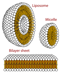
The phospholipid bilayer is formed due to the aggregation of membrane lipids in aqueous solutions.[4] Aggregation is caused by the hydrophobic effect, where hydrophobic ends come into contact with each other and are sequestered away from water.[6] This arrangement maximises hydrogen bonding between hydrophilic heads and water while minimising unfavorable contact between hydrophobic tails and water.[10] The increase in available hydrogen bonding increases the entropy of the system, creating a spontaneous process.
Function
Biological molecules are amphiphilic or amphipathic, i.e. are simultaneously hydrophobic and hydrophilic.[6] The phospholipid bilayer contains charged hydrophilic headgroups, which interact with polar water. The layers also contain hydrophobic tails, which meet with the hydrophobic tails of the complementary layer. The hydrophobic tails are usually fatty acids that differ in lengths.[10] The interactions of lipids, especially the hydrophobic tails, determine the lipid bilayer physical properties such as fluidity.
Membranes in cells typically define enclosed spaces or compartments in which cells may maintain a chemical or biochemical environment that differs from the outside. For example, the membrane around peroxisomes shields the rest of the cell from peroxides, chemicals that can be toxic to the cell, and the cell membrane separates a cell from its surrounding medium. Peroxisomes are one form of vacuole found in the cell that contain by-products of chemical reactions within the cell. Most organelles are defined by such membranes, and are called membrane-bound organelles.
Selective permeability
Probably the most important feature of a biomembrane is that it is a selectively permeable structure. This means that the size, charge, and other chemical properties of the atoms and molecules attempting to cross it will determine whether they succeed in doing so. Selective permeability is essential for effective separation of a cell or organelle from its surroundings. Biological membranes also have certain mechanical or elastic properties that allow them to change shape and move as required.
Generally, small hydrophobic molecules can readily cross phospholipid bilayers by simple diffusion.[12]
Particles that are required for cellular function but are unable to diffuse freely across a membrane enter through a membrane transport protein or are taken in by means of endocytosis, where the membrane allows for a vacuole to join onto it and push its contents into the cell. Many types of specialized plasma membranes can separate cell from external environment: apical, basolateral, presynaptic and postsynaptic ones, membranes of flagella, cilia, microvillus, filopodia and lamellipodia, the sarcolemma of muscle cells, as well as specialized myelin and dendritic spine membranes of neurons. Plasma membranes can also form different types of "supramembrane" structures such as caveolae, postsynaptic density, podosome, invadopodium, desmosome, hemidesmosome, focal adhesion, and cell junctions. These types of membranes differ in lipid and protein composition.
Distinct types of membranes also create intracellular organelles: endosome; smooth and rough endoplasmic reticulum; sarcoplasmic reticulum; Golgi apparatus; lysosome; mitochondrion (inner and outer membranes); nucleus (inner and outer membranes); peroxisome; vacuole; cytoplasmic granules; cell vesicles (phagosome, autophagosome, clathrin-coated vesicles, COPI-coated and COPII-coated vesicles) and secretory vesicles (including synaptosome, acrosomes, melanosomes, and chromaffin granules). Different types of biological membranes have diverse lipid and protein compositions. The content of membranes defines their physical and biological properties. Some components of membranes play a key role in medicine, such as the efflux pumps that pump drugs out of a cell.
Fluidity
The hydrophobic core of the phospholipid bilayer is constantly in motion because of rotations around the bonds of lipid tails.[13] Hydrophobic tails of a bilayer bend and lock together. However, because of hydrogen bonding with water, the hydrophilic head groups exhibit less movement as their rotation and mobility are constrained.[13] This results in increasing viscosity of the lipid bilayer closer to the hydrophilic heads.[6]
Below a transition temperature, a lipid bilayer loses fluidity when the highly mobile lipids exhibits less movement becoming a gel-like solid.[14] The transition temperature depends on such components of the lipid bilayer as the hydrocarbon chain length and the saturation of its fatty acids. Temperature-dependence fluidity constitutes an important physiological attribute for bacteria and cold-blooded organisms. These organisms maintain a constant fluidity by modifying membrane lipid fatty acid composition in accordance with differing temperatures.[6]
In animal cells, membrane fluidity is modulated by the inclusion of the sterol cholesterol. This molecule is present in especially large amounts in the plasma membrane, where it constitutes approximately 20% of the lipids in the membrane by weight. Because cholesterol molecules are short and rigid, they fill the spaces between neighboring phospholipid molecules left by the kinks in their unsaturated hydrocarbon tails. In this way, cholesterol tends to stiffen the bilayer, making it more rigid and less permeable.[5]
For all cells, membrane fluidity is important for many reasons. It enables membrane proteins to diffuse rapidly in the plane of the bilayer and to interact with one another, as is crucial, for example, in cell signaling. It permits membrane lipids and proteins to diffuse from sites where they are inserted into the bilayer after their synthesis to other regions of the cell. It allows membranes to fuse with one another and mix their molecules, and it ensures that membrane molecules are distributed evenly between daughter cells when a cell divides. If biological membranes were not fluid, it is hard to imagine how cells could live, grow, and reproduce.[5]
The fluidity property is at the center of the Helfrich model which allows for calculating the energy cost of an elastic deformation to the membrane.
See also
References
- ^ Murate, Motohide; Kobayashi, Toshihide (2016). "Revisiting transbilayer distribution of lipids in the plasma membrane". Chemistry and Physics of Lipids. 194: 58–71. doi:10.1016/j.chemphyslip.2015.08.009. PMID 26319805.
- ^ a b Nickels, Jonathan D.; Smith, Jeremy C.; Cheng, Xiaolin (2015). "Lateral organization, bilayer asymmetry, and inter-leaflet coupling of biological membranes". Chemistry and Physics of Lipids. 192: 87–99. doi:10.1016/j.chemphyslip.2015.07.012. PMID 26232661.
- ^ Chong, Zhi-Soon; Woo, Wei-Fen; Chng, Shu-Sin (2015-12-01). "Osmoporin OmpC forms a complex with MlaA to maintain outer membrane lipid asymmetry in Escherichia coli". Molecular Microbiology. 98 (6): 1133–1146. doi:10.1111/mmi.13202. PMID 26314242.
- ^ a b Forrest, Lucy R. (2015-01-01). "Structural Symmetry in Membrane Proteins". Annual Review of Biophysics. 44 (1): 311–337. doi:10.1146/annurev-biophys-051013-023008. PMC 5500171. PMID 26098517.
- ^ a b c d e Alberts, Bray, Hopkin, Johnson, Lewis, Raff, Roberts, Walter, Bruce, Dennis, Karen, Alexander, Julian, Martin, Keith, Peter (2010). Essential Cell Biology third edition. New York: Garland Science, Taylor & Francis Group, LLC, an informa business. p. 370. ISBN 978-0815341291.
{{cite book}}: CS1 maint: multiple names: authors list (link) - ^ a b c d e f g Voet, Donald (2012). Fundamentals of Biochemistry: Life at the Molecular Level (4 ed.). Wiley. ISBN 978-1118129180.
- ^ a b Dougherty, R. M.; Galli, C.; Ferro-Luzzi, A.; Iacono, J. M. (1987). "Lipid and phospholipid fatty acid composition of plasma, red blood cells, and platelets and how they are affected by dietary lipids: a study of normal subjects from Italy, Finland, and the USA". The American Journal of Clinical Nutrition. 45 (2): 443–455. doi:10.1093/ajcn/45.2.443. PMID 3812343. S2CID 4436467.
- ^ a b Lentz, Barry R. (2003). "Exposure of platelet membrane phosphatidylserine regulates blood coagulation". Progress in Lipid Research. 42 (5): 423–438. doi:10.1016/s0163-7827(03)00025-0. PMID 12814644.
- ^ a b Lein, Max; deRonde, Brittany M.; Sgolastra, Federica; Tew, Gregory N.; Holden, Matthew A. (2015-11-01). "Protein transport across membranes: Comparison between lysine and guanidinium-rich carriers". Biochimica et Biophysica Acta (BBA) – Biomembranes. 1848 (11, Part A): 2980–2984. doi:10.1016/j.bbamem.2015.09.004. PMC 4704449. PMID 26342679.
- ^ a b c Alberts, Bruce; Johnson, Alexander; Lewis, Julian; Raff, Martin; Roberts, Keith; Walter, Peter (2002-01-01). "The Lipid Bilayer".
{{cite journal}}: Cite journal requires|journal=(help) - ^ Daubenspeck, James M.; Jordan, David S.; Simmons, Warren; Renfrow, Matthew B.; Dybvig, Kevin (2015-11-23). "General N-and O-Linked Glycosylation of Lipoproteins in Mycoplasmas and Role of Exogenous Oligosaccharide". PLOS ONE. 10 (11): e0143362. Bibcode:2015PLoSO..1043362D. doi:10.1371/journal.pone.0143362. PMC 4657876. PMID 26599081.
- ^ Brown, Bernard (1996). Biological Membranes (PDF). London, U.K.: The Biochemical Society. p. 21. ISBN 978-0904498325. Archived from the original (PDF) on 2015-11-06. Retrieved 2014-05-01.
- ^ a b Vitrac, Heidi; MacLean, David M.; Jayaraman, Vasanthi; Bogdanov, Mikhail; Dowhan, William (2015-11-10). "Dynamic membrane protein topological switching upon changes in phospholipid environment". Proceedings of the National Academy of Sciences. 112 (45): 13874–13879. Bibcode:2015PNAS..11213874V. doi:10.1073/pnas.1512994112. PMC 4653158. PMID 26512118.
- ^ Rojko, Nejc; Anderluh, Gregor (2015-12-07). "How Lipid Membranes Affect Pore Forming Toxin Activity". Accounts of Chemical Research. 48 (12): 3073–3079. doi:10.1021/acs.accounts.5b00403. PMID 26641659.
External links
 Media related to Biological membranes at Wikimedia Commons
- Membranes at the U.S. National Library of Medicine Medical Subject Headings (MeSH)
| Authority control databases: National |
|---|