
| Ependyma | |
|---|---|
 | |
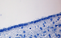 Photomicrograph of normal ependymal cells at 400× magnification in human autopsy tissue | |
| Identifiers | |
| MeSH | D004805 |
| TA98 | A14.1.00.022 |
| TA2 | 5368 |
| FMA | 242791 |
| Anatomical terminology | |
The ependyma is the thin neuroepithelial (simple columnar ciliated epithelium) lining of the ventricular system of the brain and the central canal of the spinal cord.[1] The ependyma is one of the four types of neuroglia in the central nervous system (CNS). It is involved in the production of cerebrospinal fluid (CSF), and is shown to serve as a reservoir for neuroregeneration.
YouTube Encyclopedic
-
1/5Views:80 4011 7783 99511 380285 311
-
Ependymal cells | Nervous system physiology | NCLEX-RN | Khan Academy
-
Ependymal cells
-
Ependymal tumors - Dr. Rodriguez (Hopkins) #NEUROPATH
-
Neurology | Glial Cells: Astrocytes, Oligodendrocytes, Schwann Cells, Ependymal Cells, Microglia
-
Glial Cells - Neuroanatomy Basics - Anatomy Tutorial
Transcription
In this video, I want to talk about ependymal cells. But to do so, first let me draw the brain and the spinal cord. So I'll just draw a big circle for the brain and I'll draw a long structure like this for the spinal cord. Because there are spaces inside the brain and inside the spinal cord that are full of fluid. And these spaces are connected together. And they are connected to this little skinny canal that goes down the spinal cord. And don't worry about the anatomy of this. We'll go into this in more detail in other videos. But these spaces are full of a kind of fluid called cerebral spinal fluid for the brain and the spinal cord. And the lining of these spaces is called the ependyma, which is made up of ependymal cells. The ependyma is named for a Greek word for covering. And the ependymal cells that make up the ependyma are glial cells of the central nervous system derived from neural stem cells. If we zoom in here and look at some of these ependymal cells, we'll see that they form a simple, cuboidal epithelium. Simple, meaning that they're just one layer of cells; cuboidal, meaning that they're shaped like little cubes; and epithelium, meaning they're a covering, in this case the lining of a cavity. So let's say that this is the side facing the cerebral spinal fluid, which I'll just write as "CSF" for short, for Cerebral Spinal Fluid. And that this side faces the interstitial fluid of the central nervous system, all the fluid between the cells of the brain and the spinal cord. And I'll just write "IF" as short for Interstitial Fluid. On the side of the ependymal cells facing the cerebral spinal fluid are a large number of little tiny processes called microvili, that increase the surface area of the ependymal cells on that side. They also have some processes that are a little longer, called cilia, that are these mobile, whip-like structures that kind of whip around and help move the cerebral spinal fluid around. One of the main functions of ependymal cells is to form a barrier between the cerebral spinal fluid and the interstitial fluid. So to some extent, they limit the movement of cells and large molecules between these fluid-filled spaces and the interstitial fluid of the tissue itself. Now as barriers go, the ependymal cells form a fairly leaky barrier, particularly if we were to compare it to the blood-brain barrier created by the capillaries in the central nervous system and the astrocyte end-feet. And the fact that this is a relatively leaky barrier is actually useful for medical purposes because there are areas where we can sample the cerebral spinal fluid and send it to the laboratory. And when we analyze the cerebral spinal fluid, we can often get a lot of information about what's happening in the tissue of the brain and the spinal cord because it's a relatively leaky barrier. The second major function of ependymal cells is to participate in secretion of the cerebral spinal fluid. So secreting CSF, cerebral spinal fluid. Specialized ependymal cells and capillaries form little tufts in some of these spaces in the brain. And this is actually where the cerebral spinal fluid is secreted into these spaces, so that there will be capillaries very close to the ependymal cells. And in these little tufts, fluid will be secreted across the ependymal cells to create the cerebral spinal fluid.
Structure
The ependyma is made up of ependymal cells called ependymocytes, a type of glial cell. These cells line the ventricles in the brain and the central canal of the spinal cord, which become filled with cerebrospinal fluid. These are nervous tissue cells with simple columnar shape, much like that of some mucosal epithelial cells.[2] Early monociliated ependymal cells are differentiated to multiciliated ependymal cells for their function in circulating cerebrospinal fluid.[3]
The basal membranes of these cells are characterized by tentacle-like extensions that attach to astrocytes. The apical side is covered in cilia and microvilli.[4]
Function
Cerebrospinal fluid
Lining the CSF-filled ventricles, and spinal canal, the ependymal cells play an important role in the production and regulation of CSF. Their apical surfaces are covered in a layer of cilia, which circulate CSF around the CNS.[4] Their apical surfaces are also covered with microvilli, which absorb CSF. Within the ventricles of the brain, a population of modified ependymal cells and capillaries together known as the tela choroidea form a structure called the choroid plexus, which produces the CSF.[5]
Modified tight junctions between epithelial cells control fluid release. This release allows free exchange between CSF and nervous tissue of brain and spinal cord. This is why sampling of CSF, such as through a spinal tap, provides information about the whole CNS.
Neuroregeneration
Jonas Frisén and his colleagues at the Karolinska Institute in Stockholm provided evidence that ependymal cells act as reservoir cells in the forebrain, which can be activated after stroke and as in vivo and in vitro stem cells in the spinal cord. However, these cells did not self-renew and were subsequently depleted as they generated new neurons, thus failing to satisfy the requirement for stem cells.[6][7] One study observed that ependymal cells from the lining of the lateral ventricle might be a source for cells which can be transplanted into the cochlea to reverse hearing loss.[8]
Clinical significance
Ependymoma is a tumor of the ependymal cells most commonly found in the fourth ventricle.
See also
References
- ^ "ependyma". The Free Dictionary.
- ^ Histology, a text in atlas, M. Ross 2011, 6th edition page 367
- ^ Kyrousi, C; Lygerou, Z; Taraviras, S (July 2017). "How a radial glial cell decides to become a multiciliated ependymal cell". Glia. 65 (7): 1032–1042. doi:10.1002/glia.23118. PMID 28168763. S2CID 3770948.
- ^ a b Brat, Daniel J. (2010-01-01), Perry, Arie; Brat, Daniel J. (eds.), "2 - Normal Brain Histopathology", Practical Surgical Neuropathology, New York: Churchill Livingstone, pp. 15–33, ISBN 978-0-443-06982-6, retrieved 2021-01-06
- ^ Sadler, T. (2010). Langman's medical embryology (11th ed.). Philadelphia: Lippincott William & Wilkins. p. 305. ISBN 978-0-7817-9069-7.
- ^ Johansson CB, Momma S, Clarke DL, Risling M, Lendahl U, Frisen J (1999). "Identification of a neural stem cell in the adult mammalian central nervous system". Cell. 96 (1): 25–34. doi:10.1016/S0092-8674(00)80956-3. PMID 9989494. S2CID 9658786.
- ^ Carlén M, Meletis K, Göritz C, Darsalia V, Evergren E, Tanigaki K, Amendola M, Barnabé-Heider F, Yeung MS, Naldini L, Honjo T, Kokaia Z, Shupliakov O, Cassidy RM, Lindvall O, Frisén J (2009). "Forebrain ependymal cells are Notch-dependent and generate neuroblasts and astrocytes after stroke". Nature Neuroscience. 12 (3): 259–267. doi:10.1038/nn.2268. PMID 19234458. S2CID 10479458.
- ^ "Brain cell hope for hearing loss". BBC News. 2008-12-09. Retrieved 2008-12-09.