
| Patellar ligament | |
|---|---|
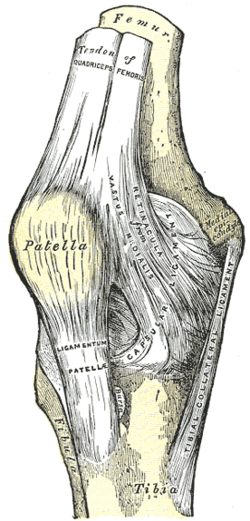 Right knee-joint. Anterior view. (Ligamentum patellae visible at bottom left, below patella.) | |
| Details | |
| From | Patella |
| To | Tuberosity of the tibia |
| Identifiers | |
| Latin | ligamentum patellae |
| MeSH | D017847 |
| TA98 | A03.6.08.015 |
| TA2 | 2622 |
| FMA | 44581 |
| Anatomical terminology | |
The patellar tendon is the distal portion of the common tendon of the quadriceps femoris, which is continued from the patella to the tibial tuberosity. It is also sometimes called the patellar ligament as it forms a bone to bone connection when the patella is fully ossified.[1]
Structure
The patellar tendon is a strong, flat ligament, which originates on the apex of the patella distally and adjoining margins of the patella and the rough depression on its posterior surface; below, it inserts on the tuberosity of the tibia; its superficial fibers are continuous over the front of the patella with those of the tendon of the quadriceps femoris. It is about 4.5 cm long in adults (range from 3 to 6 cm).[2]
The medial and lateral portions of the quadriceps tendon pass down on either side of the patella to be inserted into the upper extremity of the tibia on either side of the tuberosity; these portions merge into the capsule, as stated above, forming the medial and lateral patellar retinacula.[citation needed]
The posterior surface of the patellar tendon is separated from the synovial membrane of the joint by a large infrapatellar pad of fat, and from the tibia by a bursa.[citation needed]
Clinical significance
The patellar tendon can be injured in a patellar tendon rupture. Because tendon does not regenerate fully in humans,[3] there is a significant clinical need for research into therapies for patellar tendon rupture.
It can be used as a tissue source in the repair of other ligaments. In the event of a torn anterior cruciate ligament, the patellar tendon can be used in the rehabilitation process. In this case, the middle one third of the patellar tendon is harvested and inserted through tunnels that are drilled into the femur and tibia. The portion of the patellar tendon is then drawn through these tunnels in the bone and will be affixed to the bone via screws. The recovery process takes approximately 4–6 months upon the completion of surgery.[4] This patellar tendon method of reconstruction was traditionally the gold standard graft for anterior cruciate ligament reconstruction and is still one of the more preferred methods.[5][6][7][8]
The insertion of the patellar tendon on the tibia is the location of Osgood–Schlatter disease.
See also
Additional images
-
 Sagittal section of right knee-joint.
 -
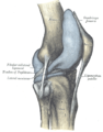 Capsule of right knee-joint (distended). Lateral aspect.
 -
 Patellar tendon. Deep dissection. Anterior view.


References
![]() This article incorporates text in the public domain from page 340 of the 20th edition of Gray's Anatomy (1918)
This article incorporates text in the public domain from page 340 of the 20th edition of Gray's Anatomy (1918)
- ^ Carreiro, Jane E. (6 November 2009). An osteopathic approach to children (2nd ed.). Edinburgh: Churchill Livingstone. ISBN 978-0-443-06738-9. OCLC 460883259.
- ^ Navali AM, Jafarabadi MA (2015). "Is There Any Correlation Between Patient Height and Patellar Tendon Length?". Arch Bone Jt Surg. 3 (2): 99–103. PMC 4468619. PMID 26110175.
- ^ James, Roshan; Kesturu, Girish; Balian, Gary; Chhabra, A. Bobby (2008-01-01). "Tendon: Biology, Biomechanics, Repair, Growth Factors, and Evolving Treatment Options". Journal of Hand Surgery. 33 (1): 102–112. doi:10.1016/j.jhsa.2007.09.007. ISSN 0363-5023. PMID 18261674.
- ^ MedlinePlus Encyclopedia: ACL reconstruction
- ^ Shaerf, Daniel A.; Pastides, Philip S.; Sarraf, Khaled M.; Willis-Owen, Charles A. (2014-01-18). "Anterior cruciate ligament reconstruction best practice: A review of graft choice". World Journal of Orthopedics. 5 (1): 23–29. doi:10.5312/wjo.v5.i1.23. PMC 3952691. PMID 24649411.
- ^ Zakko, Philip; van Eck, Carola F.; Guenther, Daniel; Irrgang, James J.; Fu, Freddie H. (2015-07-17). "Can we predict the size of frequently used autografts in ACL reconstruction?". Knee Surgery, Sports Traumatology, Arthroscopy. 25 (12): 3704–3710. doi:10.1007/s00167-015-3695-4. ISSN 1433-7347. PMID 26183732. S2CID 13441239.
- ^ http://www.aaos.org/news/aaosnow/apr12/cover1.asp[full citation needed][permanent dead link]
- ^ "Bone Patellar Bone ACL Reconstruction - Wheeless' Textbook of Orthopaedics". Retrieved 2008-10-23.
External links
- Anatomy figure: 15:01-04 at Human Anatomy Online, SUNY Downstate Medical Center - "Muscles of the anterior (extensor) compartment of the leg."
- lljoints at The Anatomy Lesson by Wesley Norman (Georgetown University) (antkneejointopenflexed)